When your doctor suspects a blockage in your urinary tract or wonders if your kidneys are shrinking, they don’t always reach for a CT scan. More often, they start with something simpler, safer, and just as powerful: renal ultrasound. It’s the go-to test for checking kidney size, spotting obstructions, and tracking changes over time-without radiation, without contrast, and without a long wait.
Why Renal Ultrasound Is the First Step
Most people think imaging means radiation, expensive machines, and long waits. But renal ultrasound breaks that mold. It uses sound waves, not X-rays, to create real-time images of your kidneys and bladder. This makes it ideal for repeated use, especially in kids, pregnant women, or anyone who needs frequent monitoring. According to the American College of Radiology, renal ultrasound is rated as "usually appropriate" for evaluating suspected urinary obstruction-higher than CT scans, which carry radiation risks. In U.S. emergency departments, it’s used in 75% of academic hospitals to quickly rule out or confirm kidney stones or blockages. Why? Because it’s fast, cheap (around $200-$500), and gives answers in minutes. The key advantage? It shows both structure and flow. You don’t just see if a kidney is enlarged-you can tell if it’s enlarged because urine is backing up. That’s called hydronephrosis, and it’s one of the clearest signs of obstruction.What Doctors Look For: Kidney Size and Shape
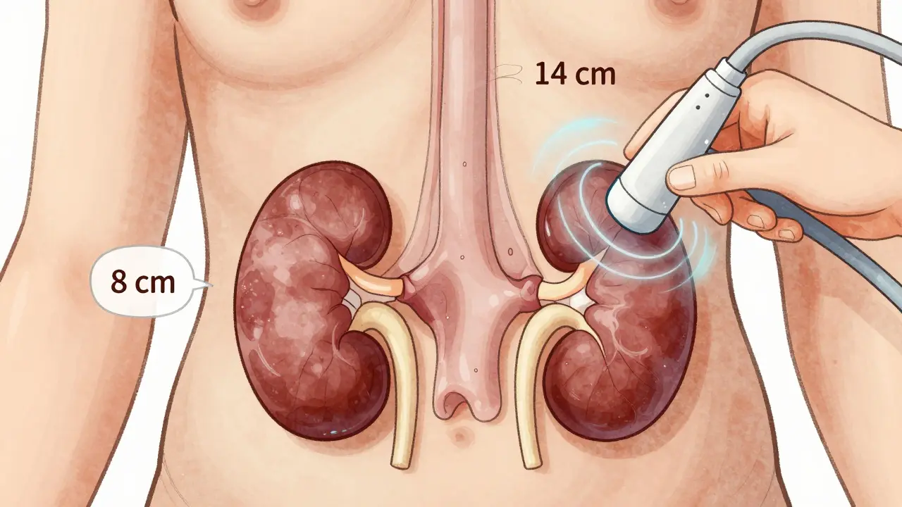
Normal adult kidneys are about 9 to 13 centimeters long. That’s roughly the size of a fist. If a kidney is smaller than 8 cm, it may be scarred or chronically damaged. If it’s larger than 14 cm, it could be swollen from obstruction, infection, or a tumor. Cortical thickness matters too. The outer layer of the kidney (the cortex) should be at least 1 cm thick. When it thins out, it often means long-term damage from high blood pressure, diabetes, or untreated blockage. A thin cortex with a dilated pelvis? That’s a red flag for chronic obstruction. Doctors measure the anteroposterior diameter of the renal pelvis-the central part where urine collects. In adults, anything over 7 mm is considered abnormal. In pregnant women, up to 10 mm is normal due to hormonal changes. But if it keeps growing week after week? That’s a sign of progressive blockage.Spotting Obstruction: Hydronephrosis and the Resistive Index
Hydronephrosis isn’t a disease-it’s a sign. It means urine isn’t draining properly. Ultrasound grades it from mild to severe:- Mild: Slight dilation of the pelvis, no change in the calyces.
- Moderate: Pelvis and calyces are widened, but the kidney still looks normal.
- Severe: The whole kidney is swollen, the cortex is thin, and the tissue looks stretched.

What Ultrasound Can’t Do (And What Can)
Ultrasound is great-but not perfect. It misses small stones. If a stone is less than 3 mm, ultrasound might not catch it. CT scans can see stones as small as 1-2 mm. So if you have intense pain and ultrasound is negative, a CT might still be needed. It also struggles in obese patients. Sound waves can’t penetrate thick layers of fat well. When BMI is over 35, the image quality drops, and doctors often switch to CT or MRI. Magnetic Resonance Urography (MRU) gives better detail of soft tissues and can show how urine moves through the system. But it costs 3-5 times more than ultrasound ($1,500-$2,500), takes longer, and still can’t reliably detect stones. Nuclear scans show kidney function but involve radiation and don’t show anatomy well. So the rule is simple: Start with ultrasound. Move to CT or MRU only if needed.New Frontiers: Elastography and AI
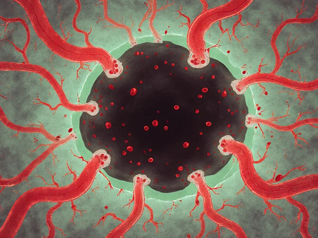
Ultrasound isn’t stuck in the 1980s. New techniques are making it even more powerful. Shear-wave elastography (SWE) measures how stiff the kidney tissue is. When urine backs up, pressure builds. That pressure makes the kidney stiffer. Studies show a clear link between increasing stiffness and worsening obstruction. This could one day let doctors measure obstruction severity without invasive tests. Super-resolution ultrasound is another breakthrough. Researchers are now able to map tiny blood vessels inside the kidney. This might help catch early kidney damage before it shows up on other tests. And AI is stepping in. Some hospitals are testing software that automatically grades hydronephrosis. Instead of relying on a radiologist’s eye, the software compares images to thousands of labeled cases. Early results are promising-accuracy is over 85%.What Happens During the Test?
There’s no special prep. You don’t need to fast. Just drink water if you’re being checked for obstruction-you want a full bladder to help see the ureters. You’ll lie on your back or side. A gel is applied to your skin. Then the technician moves a handheld probe over your flank and abdomen. It takes 15-30 minutes. You might feel slight pressure, but no pain. The technician will take measurements:- Kidney length, width, and thickness
- Cortical thickness
- Renal pelvis diameter
- Resistive index from both kidneys
- Presence of stones or masses
Who Should Get It?
Renal ultrasound is recommended for:- Patients with sudden or recurring flank pain
- People with known kidney stones
- Those with unexplained high blood pressure or abnormal blood tests (like elevated creatinine)
- Pregnant women with urinary symptoms
- Children with suspected urinary tract abnormalities
- Patients being monitored after kidney surgery
Limitations and Challenges
Even experts have trouble with it. A 2018 study found up to 20% variation in kidney size measurements between novice and experienced sonographers. That’s why training matters. The American Institute of Ultrasound in Medicine (AIUM) recommends at least 40 supervised exams before certification. Bowel gas can block the view. If you’ve eaten recently or have a lot of gas, the image may be blurry. That’s why some clinics ask you to fast for a few hours. And yes, it’s operator-dependent. If the tech isn’t trained well, they might miss a small stone or misread the RI. That’s why results should always be reviewed by a radiologist or urologist.The Bigger Picture
About 12 million renal ultrasounds are done in the U.S. each year. That’s more than any other kidney imaging test. And it’s growing. Portable ultrasound devices are now common in ERs, ICUs, and even primary care clinics. Adoption is rising because it saves time, money, and lives. The American Urological Association expects ultrasound to remain the first-line test through 2030. Even as AI and new imaging tech evolve, ultrasound’s safety, speed, and cost will keep it on top. It’s not about replacing other tools-it’s about using the right tool at the right time. For most people with suspected obstruction, that tool is still ultrasound.Can renal ultrasound detect kidney stones?
Yes, but not all of them. Ultrasound detects about 80% of kidney stones larger than 3 mm. Smaller stones (under 3 mm) are often missed. CT scans are better at finding tiny stones, but they use radiation. For most cases, especially with symptoms like pain or blood in urine, ultrasound is the first test because it’s safe and fast. If the ultrasound is negative but symptoms persist, a CT may be ordered.
Is renal ultrasound safe during pregnancy?
Yes, it’s the safest imaging option during pregnancy. Unlike CT or X-rays, ultrasound uses no radiation. It’s commonly used to check for hydronephrosis in pregnant women, which can occur due to hormonal changes and pressure from the growing uterus. If a pregnant woman has flank pain or a urinary tract infection, ultrasound is the first and often only imaging test needed.
What is hydronephrosis, and why does it matter?
Hydronephrosis is the swelling of the kidney due to a buildup of urine. It happens when something blocks the flow of urine out of the kidney-like a kidney stone, tumor, or narrowing in the ureter. Left untreated, it can damage kidney tissue over time. Ultrasound is the best way to detect and grade hydronephrosis, helping doctors decide if the blockage needs urgent treatment or can be monitored.
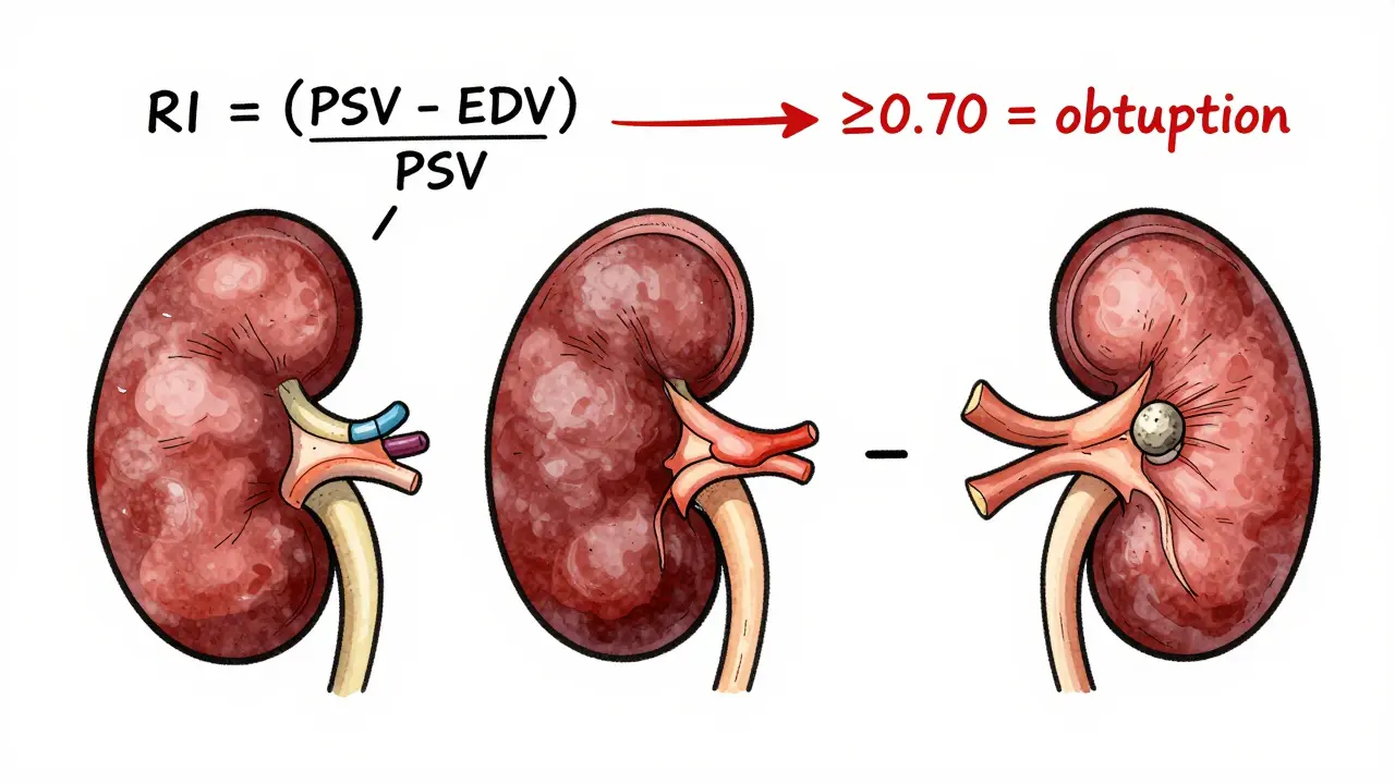
How accurate is the resistive index (RI) for diagnosing obstruction?
The resistive index is very accurate when used correctly. An RI of 0.70 or higher has been shown to detect obstruction with 86.7% sensitivity and 90% specificity, according to a 2015 study. It’s not perfect-some conditions like kidney infections or chronic disease can also raise the RI. But when combined with findings like hydronephrosis and kidney size, it becomes a powerful tool for confirming obstruction.
Do I need to prepare for a renal ultrasound?
Usually, no special preparation is needed. You can eat and drink normally. However, if you’re being checked for obstruction, you may be asked to drink water and hold your urine so your bladder is full. A full bladder helps push bowel gas out of the way and gives a clearer view of the ureters and renal pelvis. Your clinic will give you specific instructions if needed.


16 Comments
trudale hampton March 21, 2026
I've had a few renal ultrasounds over the years, and honestly, they're way less intimidating than people think. No needles, no weird prep, just some gel and a little pressure. It's wild how much info you get in 20 minutes. I wish more people knew this was the first step instead of jumping straight to CT scans.
Casey Tenney March 21, 2026
This is why we need to stop treating every kidney issue like a medical emergency. Ultrasound is the answer 90% of the time. Stop overtesting. Stop overcharging. Stop scaring people with radiation.
Allison Priole March 22, 2026
i love how ultrasound just... works. like, no fancy prep, no fasting, just drink water and chill. my cousin had hydronephrosis during her third trimester and they did an ultrasound every week. zero radiation, zero stress. she said it felt like a weird spa day. i wish all medical stuff was this chill.
Natali Shevchenko March 24, 2026
It's fascinating how something so simple-sound waves bouncing off tissue-can reveal so much about the body's internal plumbing. We've gotten so obsessed with high-tech imaging that we forget the power of the basics. Ultrasound doesn't just detect obstruction; it tells a story. The kidney isn't just big or small-it's strained, backed up, adapting. That's medicine as narrative, not just numbers.
Johny Prayogi March 24, 2026
RI ≥ 0.70 is a game changer. I've seen ER docs use this on the fly and cut diagnosis time in half. 🤘 Seriously, if you're in pain and they skip the ultrasound? That's a red flag. This tech is underused and underappreciated.
Shaun Wakashige March 25, 2026
lol i got an ultrasound last year. the tech was like 'hold your breath' and i was like 'hold it? for how long? 10 seconds? 10 minutes?'
Chris Dwyer March 27, 2026
If you're worried about kidney issues, start here. No radiation, no cost shock, no waiting. This isn't just a test-it's a smart first move. Trust the process. Your body will thank you.
shannon kozee March 29, 2026
Operator dependence is real. I've had two ultrasounds with conflicting results because one tech was rushed. Always ask if the tech is certified. AIUM-trained. Don't just accept whatever comes back.
Timothy Olcott March 29, 2026
they say ultrasound is safe but what if they're hiding something? i heard the sound waves can mess with your DNA if you do it too much. and why do they always make you drink water? are they trying to make you pee on command? 🤔
Thomas Jensen March 30, 2026
they say ultrasound is cheap but have you seen the bill? $500? for a machine that just moves a stick over your skin? they're milking us. I bet they're using the same probe on everyone. And what about the 'AI' grading? That's just a bot reading old scans. They're automating the truth. Wake up.
Paul Cuccurullo March 31, 2026
The elegance of renal ultrasound lies in its restraint. In an age of overdiagnosis, it stands as a quiet champion of clinical wisdom-measuring, observing, and refraining from the impulse to intervene without evidence. It is not merely a diagnostic tool; it is a philosophy of care.
Solomon Kindie April 2, 2026
so like if the ri is high does that mean your kidney is stressed or just that the tech was tired that day? also i think they just make up the 7mm thing because they need to sell more ct scans. i mean who even measures cortical thickness? some robot?
Nicole James April 3, 2026
Wait... so they're using ultrasound to avoid radiation... but what if the ultrasound machine itself is emitting low-level EMF? And isn't the 'resistive index' just a number they invented because they couldn't measure the real pressure? I read on a forum that the FDA once flagged these devices for 'unverified claims'... and why do they always say 'it's safe' like they're trying too hard?
Nishan Basnet April 4, 2026
In India, we use portable ultrasound in rural clinics because it's the only option. I've seen a village health worker detect hydronephrosis in a pregnant woman with no other tools. This isn't luxury tech-it's lifeline tech. The real innovation isn't AI or elastography. It's making this accessible to everyone.
Desiree LaPointe April 6, 2026
Oh, so we're celebrating a 1980s technology as if it's cutting-edge? Elastography? AI? Please. This is just the same machine with a new sticker. The real breakthrough would be if they stopped calling it 'first-line' and admitted that we're just too lazy to invest in better tools. Also, 'no radiation'-but what about the psychological radiation of being told you're fine when you're not? 😏
trudale hampton April 6, 2026
I appreciate the comment about operator dependence. I had a tech who asked me if I'd had a kidney stone before. That one question changed the whole scan. It's not just the machine-it's the person behind it. Training matters.